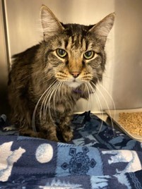
Inflammatory Bowel Disease
Species affected: felines, canines, humans, equines
Human Terminology: Inflammatory Bowel Disease, Crohn’s Disease, Ulcerative colitis
Description:
Inflammatory bowel disease (IBD) involves chronic inflammation of the gastrointestinal (GI) tract. Typical IBD symptoms in humans include severe and chronic diarrhea, abdominal pain, rectal bleeding, fatigue, and weight loss. Symptoms vary due to severity and location of inflammation. IBD in humans can be very painful and debilitating and can sometimes lead to life-threatening complications. In humans, there are two major forms of IBD: Ulcerative Colitis (UC) and Crohn’s Disease. While both are characterized by chronic inflammation of the digestive tract and share some symptoms, there are some key differences between the two. In general, they differ in which tissue layers and locations are affected as well as the continuity of the inflammation. While Crohn’s Disease can affect any area of the GI tract between the mouth and the anus, UC is limited to the colon. UC only affects the innermost lining of the colon, but Crohn’s Disease can cause inflammation in all layers of the bowel walls. UC involves continuous inflammation of the colon, meaning there are no healthy areas in between inflamed tissues; Crohn’s Disease patients may have healthy areas of the intestine in between the inflamed areas (UCLA Health).
In feline and canine patients, IBD tends to be less severe than in humans, involving chronic diarrhea and/or vomiting, often with some weight-loss, but rarely involving significant abdominal pain, rectal bleeding, or lethargy, although appetite and activity are variable. Especially in cats, the clinical picture can be influenced by concurrent disease—e.g. IBD and cholangitis (inflammation of the bile duct system) and pancreatitis (inflammation of the pancreas), called “triaditis” in cats. In dogs, location of inflammation can be quite important as they suffer from colitis (inflammation of the inner lining of the colon) more frequently than cats, where most of the disease involves inflammation in the small intestine. For this reason, canine patients are more likely to present with some blood in the stool, although not as commonly as in human patients.
In small animals, life-threatening complications from IBD are rare unless the IBD is severe and chronic enough to be categorized as a “protein-losing enteropathy” (protein loss due to compromised GI mucosa). In this case, patients are at some risk for developing blood clots or emboli or developing ascites (abdominal swelling due to the accumulation of fluid in the peritoneal cavity). Typically, IBD in small animals is often a more chronic, slowly-progressive disease than it is painful and debilitating (as seen in humans).
In horses, there is a collection of diseases that are classified as IBD, including: lymphocytic-plasmacytic enterocolitis (LPE), which is characterized by an abnormal infiltration of lymphocytes and plasma cells into the lamina propria of the intestinal wall); granulomatous enteritis (GE), which is characterized by excess infiltration of macrophages or epithelioid cells into the mucosa and submucosa of the small intestine causing the infiltrated cells to form into sheets of cells and distinctive circular granulomas; idiopathic focal eosinophilic enterocolitis (IFEE), which is characterized by eosinophilic inflammation in a focal area of the small intestine resulting in narrowing of the intestinal lumen and obstruction of flow; and multisystemic eosinophilic epitheliotropic disease (MEED). which is characterized by abnormal infiltration of eosinophils into tissues and the formation of granulomatous eosinophilic lesions in multiple organs (Merck Vet Manual).
Significant Similarities and Differences Between Affected Species:
IBD in humans, both Crohn’s and UC, often develops in the late teenage to young adult years (although it can develop at any age). In cats, IBD most often develops in middle-aged and older cats (though cats of any age can be affected). IBD in horses tends to affect younger patients, although older horses can also be affected, commonly with LPE and MEED. In all species, IBD seems to be caused by genetic abnormalities impacting the immune system and/or an inappropriate or excessive immune reaction.
IBD in humans can be very painful and debilitating and can sometimes lead to life-threatening complications. In small animals, life-threatening complications from IBD are rare unless the IBD is severe and chronic enough to be categorized as a “protein-losing enteropathy” (protein loss due to compromised GI mucosa). In this case, patients are at some risk for developing blood clots or emboli or developing ascites (abdominal swelling due to the accumulation of fluid in the peritoneal cavity). Typically, IBD in small animals is often a more chronic, slowly-progressive disease than it is painful and debilitating (as seen in humans).
Disease Etiology:
In all affected species, the cause of IBD is multifactorial and poorly understood. Previously it was thought that diet and stress were primary causes of IBD, but these are now recognized as risk factors that aggravate but do not cause IBD. (Mayo Clinic). Researchers now recognize an immune system malfunction as the main culprit leading to IBD. Similar to the autoimmune reaction seen with an allergic response, the lining of the intestine is invaded by inflammatory cells. This abnormal immune response leads to the loss or destruction of normal cell numbers and function within the tissue of the GI tract resulting in intestinal villi atrophy, which interferes with the ability to digest and absorb nutrients.
Clinical Presentation:
Significant weight loss and consistent diarrhea are typical clinical signs of IBD in animals. Rectal bleeding may also present in canine large bowel disease but is otherwise not typical and rarely seen in cats.
Since various parts of the GI tract can be affected in cats, clinical presentation may vary depending on severity and location of the inflammation. Inflammation in the colon results in chronic large bowel diarrhea and may include blood in the stool. If the stomach or areas of the small intestine are affected, the feline patient is likely to experience chronic vomiting. (Cornell Feline Health Center).
In horses, diarrhea may or may not be present. In equine patients, weight loss is primarily present in GE, LPE, and MEED. Horses with IFEE primarily present with symptoms of colic (or abdominal pain). Signs of colic in horses include pawing, excessive rolling, kicking stomach, stretching, sweating, a high pulse rate, and under-eating. This abdominal pain in horses is caused by the formation of focal inflammatory lesions resulting in the narrowing of the intestinal lumen and obstruction of ingesta flow.
Typical IBD symptoms in humans include severe and chronic diarrhea, abdominal pain, rectal bleeding, fatigue, and weight loss. Symptoms vary due to severity and location of inflammation.
Diagnosis:
In most cases, diagnosis is based on clinical symptoms. In small animals, by definition, the diagnosis of IBD requires biopsy and histopathology. Initial testing for IBD involves fecal examinations, blood testing, and intestinal imaging by ultrasound. Other signs of IBD include low serum concentration and malabsorption of carbohydrates (e.g. a failure to absorb oral glucose). Testing for B12 and folate deficiencies may be useful when diagnosing IBD in humans and animals. A low level of vitamin B12 (cobalamin) in the blood indicates a decreased ability to absorb nutrients in the distal small intestine. Decreased folate levels in the blood indicate an imbalance in the normal bacterial populations of the GI tract (VCA Hospitals).
Feline IBD has proven difficult to diagnose due to its similarity to GI lymphoma, both presenting with similar clinical signs and symptoms. For this reason, IBD in cats requires a histopathologic diagnosis. A histopathological diagnosis via microscopic examination of tissues and cells can be very useful in determining infiltrating lymphocyte cell population and origin and differentiating between IBD and GI lymphoma. Another useful diagnostic approach to decipher between IBD and GI lymphoma is clonality testing by polymerase chain reaction (PCR) to identify antigen receptor rearrangements (PARR) in the T- or B-cell receptor genes. Although these approaches are often necessary for an accurate diagnosis in feline patients, they not only require anesthesia, but, beyond that, finding a histopathologist often requires persistent efforts, expense, and a referral.
Treatment:
In both humans and animals, treatment involves the use of anti-inflammatory and immunomodulating drugs. Antibiotics may also be prescribed for anti-inflammatory effects and to “reset” the microbiome (when paired along with a probiotic). Natural supplements and remedies include dietary changes, vitamin B12, calcium, and vitamin D. IBD in small animals is often treated with a hypoallergenic or hydrolyzed diet to reduce antigenic stimulation of the immune system (fiber would be added to this diet in cases of large bowel diarrhea, i.e. colitis). In severe cases, treatment of IBD in human patients involves surgical procedures. The severity of IBD and the fact that many affected human and animal patients continue to have symptoms despite or because of current available therapies demonstrates a strong need for increased understanding and novel treatment approaches for the disease.


