Mitral Valve Disease
(myxomatous mitral valve disease, chronic mitral valve disease, degenerative mitral valve disease, mitral insufficiency)
Species affected: Humans, Canines
Brief Disease Description:
Mitral valve disease (MVD) can be congenital (a defect in the valve the patient is born with) or acquired (valve conditions that develop with aging or due to infection) and includes both mitral valve regurgitation (a leaky valve) and mitral valve stenosis (a narrow valve that cannot open). The mitral valve (left atrioventricular valve) requires the several structures including the valve leaflets, chordae tendineae, and papillary muscles to work in unison, and, if any of those structures fail or function abnormally, the valve is not able to function properly.
As dogs age, the mitral valve tends to degenerate and a significant portion of older dogs have some form of mitral valve disease. The most common outcome is mitral regurgitation, which can lead to volume overload and congestive heart failure (CHF). In humans mitral valve stenosis is usually a complication of rheumatic fever and therefore is uncommon in most developed countries.
Significant Similarities and Differences Between Affected Species:
While mitral valve regurgitation is common in dogs, mitral valve stenosis is rarely seen. Degenerative or myxomatous MVD (MMVD) is the most common cardiac condition diagnosed in dogs. Similarly, mitral valve regurgitation is the most common severe heart valve pathology in humans over 55 years of age. On post-mortem analysis, 5% to 7% of older people and about 50% of older dogs had MMVD. In both species, mitral valve degeneration seems to increase with age. Additionally, there is likely a genetic component to MMVD in both species. Small breed dogs and certain breeds such as the Cavalier King Charles Spaniel are predisposed to MMVD. An estimated 10% of all small breed geriatric dogs have MMVD. In humans, MMVD has a familial history component.
Disease Etiology:
In both dogs and humans, degeneration of the valve leads to mitral valve prolapse with subsequent regurgitation, and the lesions are characterized by thickened leaflets and elongated chordae tendineae. The risk factors for developing MMVD are similar: risk increases with age, male sex in older individuals, and family history of MMVD. The genetic component of MMVD is clear as some dog breeds are predisposed, and a familial history predisposes a person as well. Some theories suggest that individuals with generalized connective tissue abnormalities are more at risk for MMVD. This is supported by the canine model as dogs more predisposed to connective tissue disorders are also more likely to have MMVD. Research suggests that serotonin dysregulation may play a pathogenic role in MMVD in dogs, and further studies need to be conducted to determine what role, if any, serotonin dysregulation plays in human MMVD. Congenital defects of the mitral valve also occur in both humans and dogs.
Clinical Presentation:
The clinical presentation of MMVD is remarkably similar in both human and dogs. For most patients with MMVD, clinical signs early in the disease are mild or absent. In both species abnormal heart sounds such as a heart murmur can be one of the first signs of MMVD. Additional clinical signs that develop as the disease progresses can include fainting, fatigue, shortness of breath, peripheral edema, coughing, and exercise intolerance. Human patients may also report dizziness and discomfort in the chest. In both species untreated MMVD can progress to CHF, pulmonary hypertension, and pulmonary edema.
Diagnosis:
In many canine patients there is a presumptive diagnosis of MMVD if the patient is older, small breed, and has a left-sided systolic heart murmur. In human patients, a heart murmur can also indicate the possibility of MMVD. In both species an echocardiogram and chest radiographs can be performed to assess for the presence of MMVD and to monitor disease progression. Cardiac MRI is a more advanced diagnostic technique used in humans, which is rarely used for the diagnosis in dogs. There is evidence that in both species MMVD is accompanied by hypomagnesemia and platelet abnormalities.
Treatment:
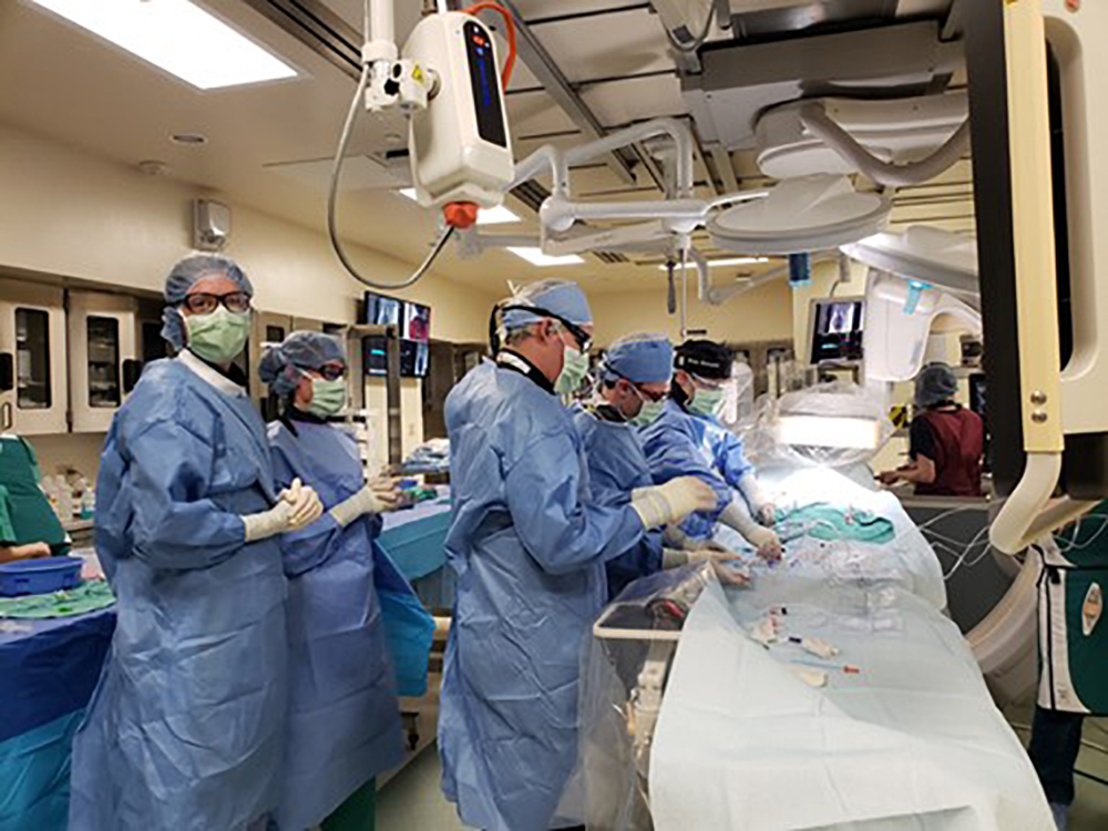
Since much of the disease is subclinical early on, treatments in both humans and dogs are often aimed at delaying disease progression and preventing the onset of CHF. Drug therapies are often used to slow the disease progression by easing the workload of the heart, improving heart muscle function, and regulating cardiac rhythm. Some treatments also include lifestyle changes such as sodium-restricted diets and restriction from extreme activities. Currently, the only way to reverse the process requires surgical intervention, such as a valve repair or replacement. These surgical procedures are common in human patients but remain rare in veterinary medicine. As interventions such as minimally-invasive transcatheter therapies become more common in humans with MMVD, there is the potential for collaboration on new technologies and techniques across species. In both species the death rate from MMVD is fairly low with an estimate 75% of dogs with MMVD dying from a different cause. However, in dogs that develop CHF, the prognosis is between ten and eighteen months. If MVD progresses to CHF in humans then it can also be a life-threatening disease.
Other Natural Animal Models:
Recent Publications/References:
Henrik D. Pedersen, Jens Häggström, Mitral valve prolapse in the dog: a model of mitral valve prolapse in man, Cardiovascular Research, Volume 47, Issue 2, August 2000, Pages 234–243, https://doi.org/10.1016/S0008-6363(00)00113-9
Fox, P. (2012). Pathology of myxomatous mitral valve disease in the dog. Journal of Veterinary Cardiology., 14(1), 103–126. https://doi.org/10.1016/j.jvc.2012.02.001
Meurs KM, Friedenberg SG, Williams B, et al. Evaluation of genes associated with human myxomatous mitral valve disease in dogs with familial myxomatous mitral valve degeneration. Vet J. 2018;232:16-19. doi:10.1016/j.tvjl.2017.12.002
Connell PS, Han RI, Grande-Allen KJ. Differentiating the aging of the mitral valve from human and canine myxomatous degeneration. J Vet Cardiol. 2012;14(1):31-45. doi:10.1016/j.jvc.2011.11.003
Oyama MA, Elliott C, Loughran KA, et al. Comparative pathology of human and canine myxomatous mitral valve degeneration: 5HT and TGF-β mechanisms. Cardiovasc Pathol. 2020;46:107196. doi:10.1016/j.carpath.2019.107196
Aupperle H, Disatian S. Pathology, protein expression and signaling in myxomatous mitral valve degeneration: comparison of dogs and humans. J Vet Cardiol. 2012;14(1):59-71. doi:10.1016/j.jvc.2012.01.005
Orton EC, Lacerda CM, MacLea HB. Signaling pathways in mitral valve degeneration. J Vet Cardiol. 2012;14(1):7-17. doi:10.1016/j.jvc.2011.12.001
Disatian S, Ehrhart EJ 3rd, Zimmerman S, Orton EC. Interstitial cells from dogs with naturally occurring myxomatous mitral valve disease undergo phenotype transformation. J Heart Valve Dis. 2008;17(4):402-412.
Griffiths LG, Orton EC, Boon JA. Evaluation of techniques and outcomes of mitral valve repair in dogs. J Am Vet Med Assoc. 2004;224(12):1941-1945. doi:10.2460/javma.2004.224.1941
Monnet E, Orton EC, Gaynor J, Boon J, Peterson D, Guadagnoli M. Diagnosis and surgical repair of partial atrioventricular septal defects in two dogs. J Am Vet Med Assoc. 1997;211(5):569-572.


